Available Microscopic Capabilities
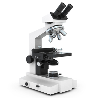
- Variety of Optical Light Microscopes
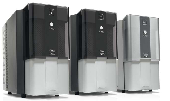
- SEM – Scanning Electron Microscopy
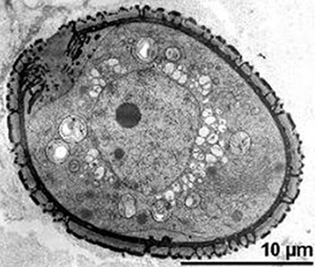
- TEM – Transmission Electron Microscopy
- EDS – Energy Dispersive X-Ray Spectroscopy
The classification and identification of small particles begins with observation of the particle through a microscope using a combination of transmitted and reflected light. Particles photographed through the microscope can be measured and displayed for a report or an advertisement especially when the particles’ harbor unique characteristics. We have available several different variants of light microscopes to meet the needed magnifications for observation of your sample.


When a second test is needed to confirm an observation made by light microscopy, or when a particle is opaque, the particle can be imaged and analyzed using electrons instead of light. The immediate difference is that the images produced by the SEM are gray scale. The images reveal a depth of field and detail that is superior to light microscopy with an added bonus that as electrons are bombarding the sample, x-rays are produced that are representative of the elements present in the sample. By adding an energy dispersive x-ray spectrometer (EDS) it enables the ability to determine the elements present in the sample while imaging the sample.
Particles too small to be analyzed and imaged by light microscopy or scanning electron microscopy must be observed and analyzed in the transmission electron microscope. For thin samples or samples that can be made thin, TEM imaging techniques can reveal the crystalline structure of the particle as well as its elemental composition (EDS). Clays, pigment particles, thin films and other nanometer-sized particles can be analyzed and identified in the TEM. Some materials such as thin mineral fibers, clay particles, and soot need very little preparation to be analyzed. Other materials require considerable sample preparation prior to analysis.

Particles that are plastic (easily deformed) can be characterized using a microscope that uses reflected and transmitted infrared light. Polymeric materials that need to be characterized and identified can be prepared for FTIR. The resulting infrared spectrum can be compared to thousands of reference spectra to determine the type of polymer. Manufacturing facilities that occasionally have undesirable particles appear in their finished products will keep a reference library of infrared spectra that identify the materials used in their processes to aid in the characterization of customer returns.
The characterization of surfaces requires a slightly different approach using a microscope that can measure surface roughness and provide a three-dimensional image of the surface. This microscope utilizes reflected white light while scanning through focus to create a three-dimensional data set from which surface roughness parameters can be calculated. Unlike stylus profilometry, SWLIM is a non-contact technique with little or no sample preparation required.
You can see how this popup was set up in our step-by-step guide: https://wppopupmaker.com/guides/auto-opening-announcement-popups/
You can see how this popup was set up in our step-by-step guide: https://wppopupmaker.com/guides/auto-opening-announcement-popups/
You can see how this popup was set up in our step-by-step guide: https://wppopupmaker.com/guides/auto-opening-announcement-popups/
"*" indicates required fields
"*" indicates required fields
"*" indicates required fields
"*" indicates required fields
"*" indicates required fields
"*" indicates required fields
"*" indicates required fields
"*" indicates required fields
"*" indicates required fields
"*" indicates required fields
"*" indicates required fields
"*" indicates required fields
"*" indicates required fields
"*" indicates required fields
"*" indicates required fields
"*" indicates required fields
"*" indicates required fields
"*" indicates required fields
"*" indicates required fields
"*" indicates required fields
"*" indicates required fields
"*" indicates required fields